Leading expert in structural heart interventions, Dr. Francesco Maisano, MD, explains how advanced cardiac imaging with CT, MRI, and echocardiography is fundamental for planning complex heart valve treatments. He details the shift from a single-modality approach to a sophisticated, multi-modality diagnostic strategy that enables personalized procedural planning, simulation, and improved patient outcomes for aortic stenosis, mitral regurgitation, and tricuspid valve disease.
More from Heart Valve [Aortic, Mitral, Tricuspid]

More from Diagnostic Detectives Network

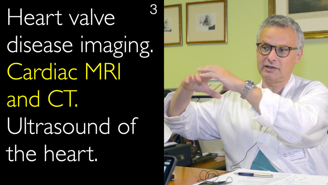
Advanced Cardiac Imaging for Heart Valve Disease Treatment Planning
Jump To Section
- Multimodality Imaging Revolution
- Cardiac CT as Standard Practice
- Echocardiography as a Mainstay
- Cardiac MRI's Emerging Role
- Procedure-Specific Imaging Pathways
- The Future of Simulation and Prediction
- Full Transcript
Multimodality Imaging Revolution
Dr. Francesco Maisano, MD, emphasizes that diagnostic imaging innovation has progressed in parallel with the development of new medical devices for heart disease. He explains to Dr. Anton Titov, MD, that modern cardiac care relies on an integrated, multimodality imaging approach. This strategy is fundamental not only for planning complex heart valve and coronary artery treatments but also for guiding the procedures themselves, whether they are open-heart surgeries or percutaneous transcatheter interventions.
The classical diagnostic pathway, which was mainly driven by echocardiography and angiography, is being replaced. Patients now often undergo two or three different imaging modalities to establish a precise treatment indication and develop the optimal strategy. This comprehensive approach allows physicians to select the correct medical device for endovascular procedures and to meticulously plan even the most complex open-heart operations.
Cardiac CT as Standard Practice
Cardiac computed tomography (CT) has become a standard of care in the planning of structural heart interventions. Dr. Francesco Maisano, MD, states that in his practice, a heart CT scan is performed on every patient scheduled for a procedure. He describes it as a "one-stop shop" that provides a wealth of critical information, including detailed coronary artery anatomy, access points, and the extent of calcification.
Beyond its primary purpose, cardiac CT frequently reveals unexpected findings, such as tumors, that require medical attention. This makes the scan a necessary step for any patient indicated for a structural heart disease intervention. The data from a CT scan is indispensable for answering key clinical questions, such as determining the severity of aortic stenosis and deciding between surgical aortic valve replacement and a transcatheter aortic valve implantation (TAVI) procedure.
Echocardiography as a Mainstay
Despite the rise of advanced imaging, Doppler echocardiography remains the mainstay of initial heart disease diagnosis and screening. Dr. Francesco Maisano, MD, highlights its enduring importance due to its simplicity, wide availability, and bedside capability. Echocardiography serves as the first diagnostic step, providing essential functional and anatomical data on heart valves and chambers.
The technology continues to evolve, becoming more sophisticated each year with the integration of 3D imaging and predictive algorithms. For certain procedures, echocardiography provides all the necessary data. Transesophageal echocardiography (TEE) is particularly vital for intraprocedural guidance during mitral valve interventions, offering real-time imaging that is crucial for a successful outcome.
Cardiac MRI's Emerging Role
Cardiac magnetic resonance imaging (MRI) is emerging as a powerful functional diagnostic test in cardiology. Dr. Francesco Maisano, MD, explains to Dr. Anton Titov, MD, that cardiac MRI is particularly valuable for identifying the right timing for treatment and clarifying indications, especially in complex heart failure patients. Its ability to assess intracardiac fluid flow and fluid dynamics represents a new frontier in understanding cardiac pathophysiology.
While the clinical application of this flow dynamic data is still evolving, Dr. Francesco Maisano, MD, is confident it will become a fundamental piece of information for planning future cardiac treatments. Cardiac MRI is gaining importance for patients presenting with complex conditions like low-flow, low-gradient aortic stenosis or functional mitral and tricuspid regurgitation, where more sophisticated functional data is required beyond basic anatomy.
Procedure-Specific Imaging Pathways
Every cardiac pathology requires a tailored multimodality imaging package designed to answer specific clinical questions. Dr. Francesco Maisano, MD, illustrates this by comparing mitral and tricuspid valve procedures. Although both are atrioventricular valves, their imaging protocols differ significantly; a CT scan for a tricuspid valve uses a different acquisition timing to capture contrast in the right heart chambers.
The choice of intraprocedural guidance also varies. While transesophageal echocardiography is perfect for mitral valve work, its utility is limited for tricuspid valve assessment, leading physicians to explore alternatives like intracardiac echocardiography. The diagnostic pathway is simpler for straightforward aortic stenosis, often requiring only echocardiography and CT. In contrast, planning for heart failure surgeries or interventions for complex valvular disease demands a more extensive battery of diagnostic tests to gather sufficient functional data.
The Future of Simulation and Prediction
The field of cardiac diagnostic imaging is moving toward predictive simulation, making the planning phase as critical as the operation itself. Dr. Francesco Maisano, MD, describes how physicians now use simulators to rehearse complex procedures and predict the effect of an implant on cardiac physiology before a patient ever enters the operating room.
This technology can forecast outcomes like flow dynamics after coronary angioplasty, the distribution of calcium post-TAVI, or the risk of complications such as perivalvular leak or heart block. Dr. Maisano tells Dr. Anton Titov, MD, that the application of finite element models and other advanced technologies is reducing improvisation in cardiac care. The profession is increasingly focused on meticulous planning, standardization, and ultimately, delivering truly personalized medicine by selecting the ideal treatment solution for each individual patient.
Full Transcript
Dr. Anton Titov, MD: Heart valve and coronary artery treatment procedures are very complicated. Open heart surgery and percutaneous transcatheter procedures are sometimes combined. The correct method of treatment has to be chosen and adjusted for each patient with heart disease. What diagnostic imaging studies are used to plan heart valve treatments today?
Dr. Francesco Maisano, MD: First of all, diagnostic imaging is as important in the evolution of new heart disease treatments as the development of new technologies and new devices. There has been a parallel pathway in diagnostic imaging and treatment innovation. On one side, we have been innovating in implantable devices. On the other side, we have been innovating and using innovation in diagnostic imaging.
Diagnostic imaging is fundamental not only for the planning of heart disease treatment but also for the conduction of treatment procedures. Endovascular structural intervention is guided by diagnostic imaging.
As far as planning, we have learned that in structural interventions today, we have massive use of preprocedural diagnostic imaging. Specifically, we use a lot of CT scans. Heart CT is becoming standard practice. In my practice, cardiac CT is standard not only for cardiac endovascular procedures but also for open cardiac surgical procedures.
In every patient I treat, I like to have a CT scan of the heart for many reasons. First of all, it is a one-stop shop for all the information about coronary artery access, coronary artery anatomy, coronary calcifications, and everything. Second, cardiac CT scan shows in a significant number of patients some unexpected findings. Unfortunately, sometimes we find some tumors or other problems that need medical attention.
In general, a cardiac CT scan is becoming a necessary step for each patient who is indicated for a structural heart disease intervention. But obviously, the mainstay of our decision-making is Doppler echocardiography. This diagnostic test remains today. Echocardiography is the first step for diagnosis of heart disease and for screening.
Echocardiography is very simple to perform. It is widely available and can be done at the bedside. It is becoming every year more and more sophisticated. There are 3D technologies and all kinds of predictive algorithms.
Cardiac MRI is also emerging as an interesting diagnostic test. It's a functional diagnostic test. It can also be used for identifying the right timing of treatment and indications for treatment.
In general, we see the transition away from the classical heart disease diagnostic pathway, which was mainly driven by echocardiography and angiography. We move towards a more integrated multimodality diagnostic imaging approach. We have patients undergoing two or three different diagnostic imaging modalities to set up the indication for treatment.
We develop the strategy to select the correct medical device in case of cardiac endovascular procedures. Even in the case of open-heart surgery, we use multimodality diagnostic imaging to plan the procedure and prepare for it.
In some cases, we even have simulators of the planned intervention. We are simulating the effect of an implant on the heart. We have simulators where sometimes you can even train yourself before the intervention for complex cardiac procedures.
Cardiac diagnostic imaging is an incredible field. It is becoming as important as the heart operation itself. Because through diagnostic imaging today, we can really predict what is going to happen with these heart treatment procedures.
The next step is going to be the application of finite element models and other technologies to predict the effect of an implant on cardiac physiology and structure. Imagine today you can predict the effect of a coronary artery angioplasty on the flow dynamics of the coronary arteries. Or you can predict what happens with a coronary artery bypass grafting in one lesion.
You can predict how the calcium will distribute after you implant a TAVI. You can predict if there will be some atrioventricular block or if there will be some perivalvular leak. This can all be simulated with current diagnostic imaging technologies.
They are becoming more and more available. This means that our profession is becoming less and less about improvisation and more and more about planning, standardization, and selecting the ideal treatment solution for the individual patient. This is individualized medicine, truly personalized medicine for all these structural cardiac treatment interventions.
Dr. Anton Titov, MD: That's very important! Obviously, the CT scan of the heart also follows echocardiography, transthoracic versus transesophageal echocardiography. It helps to plan the heart treatment procedure. Are there any particular differences in diagnostic tests for people undergoing a mitral valve or aortic valve intervention versus patients undergoing perhaps heart failure surgery or other procedures? Is there a particular procedure-planning path that you use for different groups of patients?
Dr. Francesco Maisano, MD: Every pathology has a different multimodality diagnostic imaging package. It depends on the questions you need to answer. Let's make an example of a patient undergoing mitral valve or tricuspid valve surgery. These valves look very similar. They are both atrioventricular heart valves.
But imagine, first of all, the CT scan for a mitral valve is done by a different protocol than the CT scan for tricuspid valve. The timing of the image acquisition is different because you need to catch the dye in the right heart chamber. In the right side of the heart, if you want to do a CT scan for the tricuspid valve, we really need to measure the right heart function with right heart catheterization.
Right heart catheterization is very rarely performed for mitral valve interventions. For instance, intraprocedural guidance is done by transesophageal echocardiography. It is perfect for my job. It doesn't work so well for the tricuspid valve assessment. We still use it, but we are looking for alternatives.
We are looking for intracardiac echocardiography, for instance, because of the quality of the image that you get from transesophageal echocardiography in this particular application. It also depends very much on the heart treatment procedure we plan to do.
There are some procedures that are totally based on echocardiography data. For some other heart disease treatment procedures, we rely more on fluoroscopy. In some occasions, we even use fusion imaging. For the diagnostic phase before the cardiac interventions, it very much depends on the questions we are asking.
Let's say you have a patient with aortic stenosis who will undergo an aortic valve replacement or TAVI. This patient's situation usually has two main questions. The number one question is this: is the aortic stenosis severe, yes or no? The second question is, what should I do? Should I do surgery or a TAVI?
These are two questions that can be answered by echocardiography and by a heart CT scan. With quick answers to these two big questions, you answer 99% of the problems needed to make the decision how to treat a patient with aortic valve stenosis. Then you can check the coronary arteries, usually by a CT scan and many other tests. At the end of the day, you don't need so many diagnostic imaging steps.
The situation is very different if you talk about heart failure patients that require structural surgical intervention. These are patients with tricuspid regurgitation, mitral regurgitation, low flow or low gradient across the aortic valve. In these cases, the diagnostic pathway is a bit more sophisticated.
You need more diagnostic examinations and more functional data. In this field, cardiac MRI is not yet well established, but it's growing in importance. I think we start learning what to do with cardiac MRI. Cardiac MRI is also today focusing on intracardiac fluid flow and fluid dynamics, which is an emerging field.
We see this information but we don't know what to do with that data. We see the flow, how the blood moves in the chamber, how pathology is affecting these flow dynamics, but we don't know yet how to use them. I'm pretty sure that this is going to be one of the fundamental pieces of information that we want to achieve to plan cardiac treatment procedures.