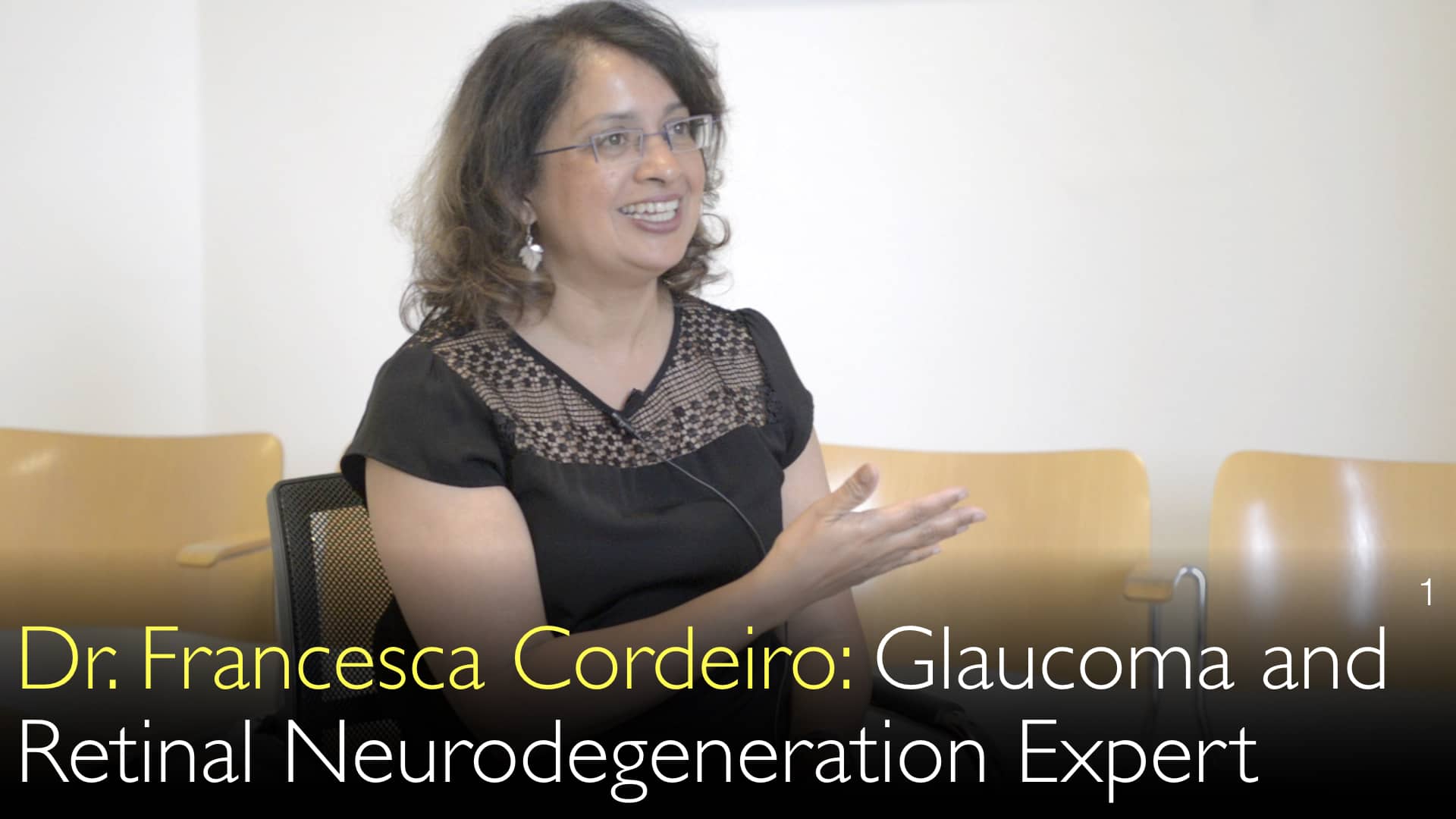
緑内障診断・治療の世界的権威であるFrancesca Cordeiro医学博士は、光干渉断層計(OCT)や視野検査といった現代的な診断技術が、この「沈黙の疾患」の早期発見に不可欠であると述べています。博士は、OCT画像解析によって、患者が自覚的な視力低下を感じるずっと前から網膜神経線維層の菲薄化を検出できると指摘。これにより、視神経への不可逆的な損傷を防ぐ早期介入が可能となり、OCTがその決定的な役割を果たすと詳しく説明しています。
緑内障の高度診断:OCTと視野検査の解説
セクションへ移動
- 緑内障とは?早期診断が重要な理由
- 眼圧測定を超えて:緑内障診断の多角的アプローチ
- 光干渉断層計(OCT):画期的な診断ツール
- 視野検査:機能的視覚障害の測定
- OCT対視野検査:どちらが早期に緑内障を検出するか?
- 診断基準値の設定が極めて重要である理由
- 経時的な緑内障進行のモニタリング
緑内障とは?早期診断が重要な理由
緑内障は、多くの場合自覚症状なく視神経に進行性の損傷を与える一群の眼疾患です。フランチェスカ・コルデイロ医学博士は、緑内障が「沈黙の病」であると強調します。つまり、患者は通常、重大かつ不可逆的な神経損傷が生じるまで視覚障害に気づきません。この無自覚な進行のため、客観的な診断検査による早期発見と失明予防のための治療が不可欠です。
眼圧測定を超えて:緑内障診断の多角的アプローチ
緑内障の診断は、眼圧測定だけでは不十分です。アントン・チトフ医学博士は、包括的な診断には複数の検査が必要だと指摘します。フランチェスカ・コルデイロ医学博士も、検査の組み合わせの重要性を確認しています。この多角的アプローチには、検眼鏡による眼底観察、視野測定、OCTなどの高度な画像診断が含まれます。これらの検査結果を総合的に評価することで、確実な緑内障診断が可能となります。
光干渉断層計(OCT):画期的な診断ツール
光干渉断層計(OCT)は、緑内障診断における主要な非侵襲的画像検査です。フランチェスカ・コルデイロ医学博士は、OCTを「非組織学的な眼の光学的断層像」を提供する技術と表現し、網膜層の詳細な断面画像を得られると説明します。この検査は特に、網膜神経線維層と網膜神経節細胞層に焦点を当てます。緑内障ではこれらの層が菲薄化し、OCTはこの構造変化を高精度で検出できます。
視野検査:機能的視覚障害の測定
視野検査(ペリメトリー)は、緑内障診断のもう一つの基盤となる検査です。この検査では、緑内障で最初に障害されやすい周辺視野を含む、患者の全視野をマッピングします。アントン・チトフ医学博士は、診断プロセスにおけるその重要性について論じています。ゴールドスタンダードである一方、フランチェスカ・コルデイロ医学博士は、視野欠損が現れるまでには相当量の神経細胞の損失が必要なため、これは疾患の後期徴候であると指摘します。
OCT対視野検査:どちらが早期に緑内障を検出するか?
緑内障医療における重要な進歩は、検出可能な変化の順序の理解です。フランチェスカ・コルデイロ医学博士は、OCTが視野検査で変化が認められる前に、網膜神経線維層の菲薄化を識別できると説明します。これにより、OCTは将来の視力喪失の強力な予測因子となります。機能的視覚が失われる数年前にこの構造的変化を確認できる能力は、はるかに早期の介入と治療を可能にします。
診断基準値の設定が極めて重要である理由
リスクのある個人にとって、基準値の設定は極めて重要です。アントン・チトフ医学博士は、正常視力で自覚症状のない人でも、参照点を作成するためにこれらの検査が必要であると強調します。この基準値により、眼科医は将来の検査結果と比較し、早期緑内障を示唆する微妙な変化を特定できます。この段階での早期発見は、長期的な視機能保存の最良の戦略です。
経時的な緑内障進行のモニタリング
OCTと視野検査の真の価値は、初期診断を超えて継続的な経過観察にあります。フランチェスカ・コルデイロ医学博士は、これらの検査が時間の経過とともに周辺視野の変化が進行しているかどうかを確認するために使用されると強調します。逐次のOCTスキャンと視野マップを比較することで、医師は疾患の進行速度を客観的に測定し、視神経へのさらなる損傷を遅らせたり停止させたりするために治療計画を調整できます。
全文
OCT(光干渉断層計)は緑内障診断の重要な検査です。OCTは緑内障の進行過程の指標として網膜神経線維を観察します。視野検査も重要ですが、網膜の多くの神経細胞が死滅した時点で現れる緑内障の後期徴候です。
アントン・チトフ医学博士: 高眼圧(眼内圧)は緑内障の唯一の検査ではありません。検眼鏡による眼底観察を含む複数の検査があります。緑内障診断は視野測定にも依存します。
フランチェスカ・コルデイロ医学博士: はい。すべての診断検査を総合することで、患者が無自覚性緑内障であるという診断を確定できます。無自覚であるため、眼疾患の有無を客観的に測定する方法が必要です。
アントン・チトフ医学博士: 我々が次第に認識してきたことの一つは、緑内障診断のゴールドスタンダードが視野検査であるにもかかわらず、視野が影響を受けるまでにはかなり多くの眼神経細胞が失われなければならないということです。そこでここ数年で発展したことの一つが、視野欠損の進行を確認する能力です。
フランチェスカ・コルデイロ医学博士: 我々はOCT(光干渉断層計)測定を行うことができます。光干渉断層計は網膜神経線維を観察します。視神経の変化は、緑内障性眼疾患があることを示す指標です。
時間の経過とともに周辺視野の変化が進行しているかどうかを確認することは、視野検査の実施の重要性を強調します。たとえ正常視力で視覚に関する自覚症状がなくても、視力基準値を設定することが極めて重要です。
はい、その通りです。しかし明らかになってきたことの一つは、眼底画像化の能力です。これは過去10年間で向上しました。
大きな進歩の一つが、光干渉断層計(OCT)の使用です。光干渉断層計は、ほとんど非組織学的な眼の光学的断層像を作成しています。網膜は多くの異なる神経細胞層で構成されています。これらはすべて特殊化されていますが、緑内障性眼疾患ではそれぞれ異なる損傷を受けます。
フランチェスカ・コルデイロ医学博士: 緑内障では、表層は網膜神経節細胞で構成されています。この表層が、視野検査に変化が現れる前でさえ菲薄化することがわかっています。その網膜層の厚みの減少の変化を探すことができます。
アントン・チトフ医学博士: それ自体が、将来視野が失われることを示す指標です。それは緑内障の徴候です。


